Advanced Imaging & Diagnostics
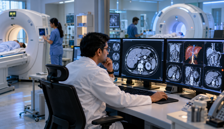
Many liver conditions are silent, and diagnosis often depends on imaging. Whether it is detecting fatty liver, measuring scarring, finding a small tumour or planning a transplant, the quality of imaging and the expertise of the radiologist shape the treatment. Our Advanced Imaging & Diagnostics department is set up to provide accurate, reliable results with minimal delay.
Imaging Services
Ultrasound and Doppler
- Abdominal ultrasound for liver, gallbladder, bile ducts, pancreas, kidneys and spleen
- Doppler studies of the portal vein, hepatic veins and hepatic artery
- Screening ultrasound every six months for patients with cirrhosis or chronic hepatitis B
- Ultrasound-guided biopsies and drainage procedures
- Contrast-enhanced ultrasound (CEUS) to characterise liver lesions in selected patients
FibroScan (Transient Elastography)
- Measures liver stiffness (fibrosis) and controlled attenuation parameter (fat) without a needle
- Takes only a few minutes and involves no radiation
- Used to diagnose and follow fatty liver, hepatitis and cirrhosis
CT Scan
- Multiphase CT for detection and characterisation of liver tumours
- CT angiography for liver vessels, donor evaluation and transplant planning
- Liver volumetry and 3D reconstruction for surgical planning
- Assessment of abdominal emergencies and complications
MRI and MRCP
- MRI of the liver with or without liver-specific contrast for lesion characterisation
- MRCP for bile ducts and pancreatic ducts, without invasive procedures
- Quantification of liver fat and iron in selected protocols
- Assessment of polycystic disease, vascular conditions and complex lesions
Other imaging
- Chest and abdominal X-ray
- Echocardiography and bubble-contrast echo for cardiac and lung evaluation in transplant candidates
- Nuclear medicine and PET-CT for selected cancers, where available
- Bone density scans for patients with cholestatic and advanced liver disease
Diagnostic Support Beyond Imaging
- Endoscopy and EUS: Direct visualisation and tissue sampling
- Liver biopsy: Percutaneous or transjugular, guided by imaging and performed with safety checks
- Pulmonary function tests: For assessing lung health before surgery and transplant
- Cardiac evaluation: ECG, stress testing and echocardiography
- Coordination with pathology and laboratory services for integrated diagnosis
Specialised Liver Imaging Applications
- Liver cancer screening and diagnosis: Multiphase CT and MRI can confirm hepatocellular carcinoma without biopsy in cirrhotic patients, using standard imaging criteria.
- Transplant assessment: CT or MR angiography maps the vessels and bile ducts of donors and recipients, while volumetry ensures the graft size is adequate.
- Post-transplant surveillance: Doppler ultrasound checks hepatic artery, portal vein and hepatic veins for narrowing or clots. MRCP helps detect bile duct problems.
- Portal hypertension assessment: Ultrasound, CT and Doppler show varices, spleen size, portal vein thrombosis and collateral vessels.
- Fatty liver quantification: FibroScan and MRI-based methods measure fat and fibrosis more accurately than ultrasound alone.
Radiation Safety and Contrast Use
- Imaging protocols follow the principle of keeping radiation as low as reasonably achievable.
- Ultrasound, FibroScan and MRI use no ionising radiation.
- Kidney function and allergy history are checked before contrast is given.
- Patients with cirrhosis and kidney risk receive careful hydration and contrast choices.
- Pregnancy status is checked before relevant tests.
Preparation for Common Tests
- Ultrasound: Fasting for 6 to 8 hours and a full bladder if the pelvis is also being examined
- FibroScan: Fasting for at least 2 to 3 hours is generally advised
- CT and MRI: Fasting as instructed, removal of metal objects, and information about implants, pacemakers and claustrophobia
- MRCP: Fasting for several hours
- Medicines: Continue usual medicines unless told otherwise, and inform staff about diabetes tablets, especially metformin, before contrast studies
The team gives written instructions at the time of booking.
Reporting and Communication
Radiologists with experience in hepatobiliary and transplant imaging review each study. Reports use standardised terminology and, where relevant, recognised systems such as LI-RADS for liver lesions. Urgent findings are communicated directly to treating doctors, and complex cases are discussed in joint meetings with hepatologists, surgeons and interventional radiologists.
Integration With Care
Images and reports are available to the treating team for quick decisions. This helps in:
- Faster diagnosis in OPD and emergency
- Planning surgeries, transplants and interventional procedures
- Monitoring treatment response in cancer and cirrhosis
- Screening high-risk patients on schedule
Patient Comfort
Our staff explain each test, reassure anxious patients and support children and elderly patients. Comfortable waiting areas, clear signage and helpful staff reduce stress.
Understanding Your Report
Patients often feel worried when reading medical terms. Our doctors go through reports in simple language and explain which findings matter and what to do next. Please bring old scans for comparison, since changes over time are often more informative than a single image.
Book Your Investigation
For imaging or diagnostic tests related to liver, bile duct or digestive conditions, contact LiverMitra to book an appointment with our diagnostics team.